Multiple sclerosis is a neuroimmune disease with activated microglia playing a key role in the disease. The disease is associated with a loss of myelin that contributes to the loss of function. The immune system, T-cells in particular, play a key role and antibody therapies directed at T-cells are used to treat the disease.
16x20” multimedia
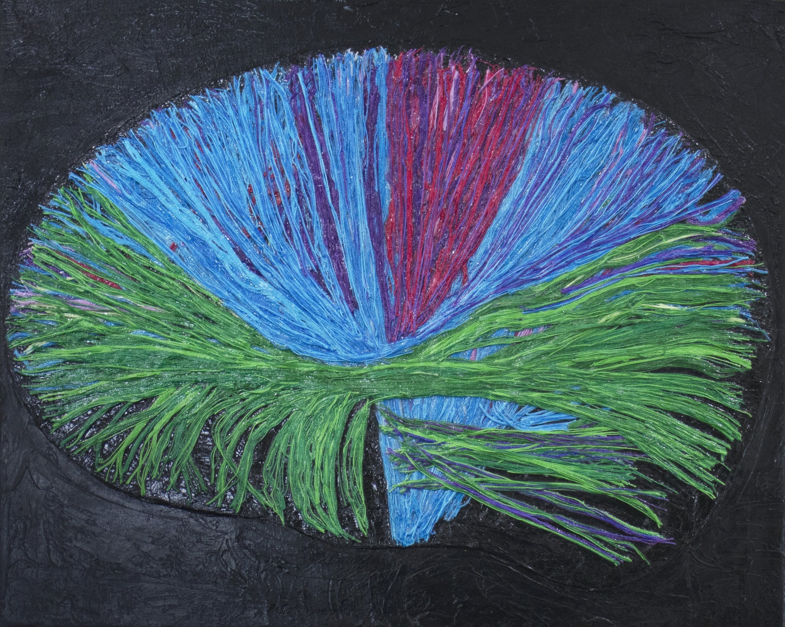
Diffusion tensor imaging (DTI) is a form of MR imaging technique that is used to look at white matter in the brain. All based on the movement of water. #Brainart #sciart #slukaart #brainimaging #neuroart.

SOLD
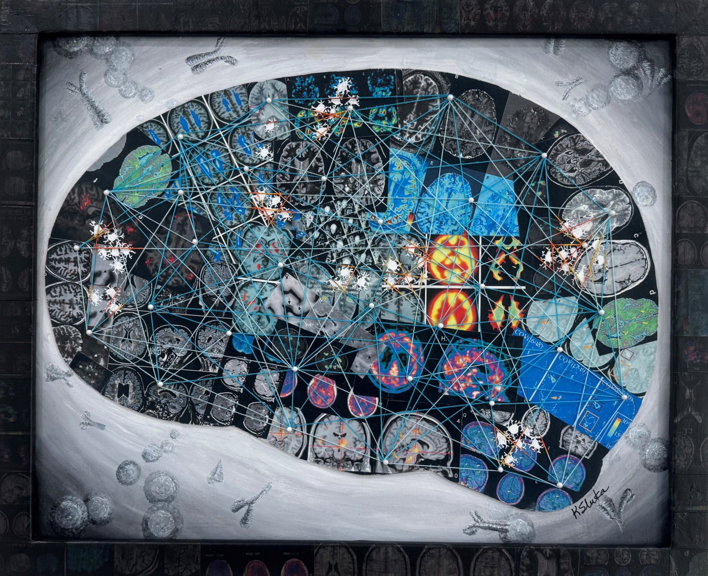
Brain imaging studies have transformed our understanding of how pain is processed in the brain. This multimedia collage uses brain images and text from published studies by our favorite pain scientists.
Multimedia, 16x20”
SOLD
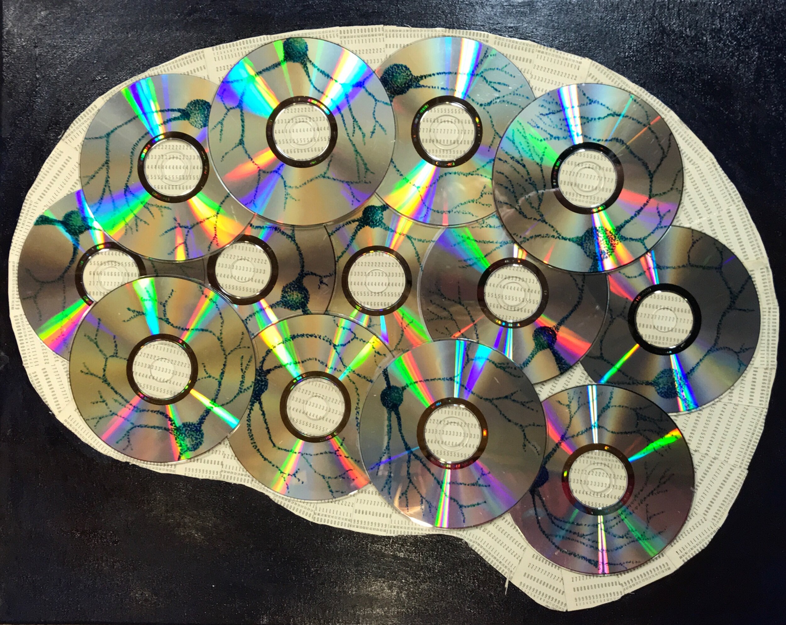
The brain represents the computer-like storage of data in many forms by neurons. This brain is collaged from old CDs. I drew neurons on each and collaged the background from IBM punch cards.
16x20 Multimedia collage and acrylic painting
SOLD
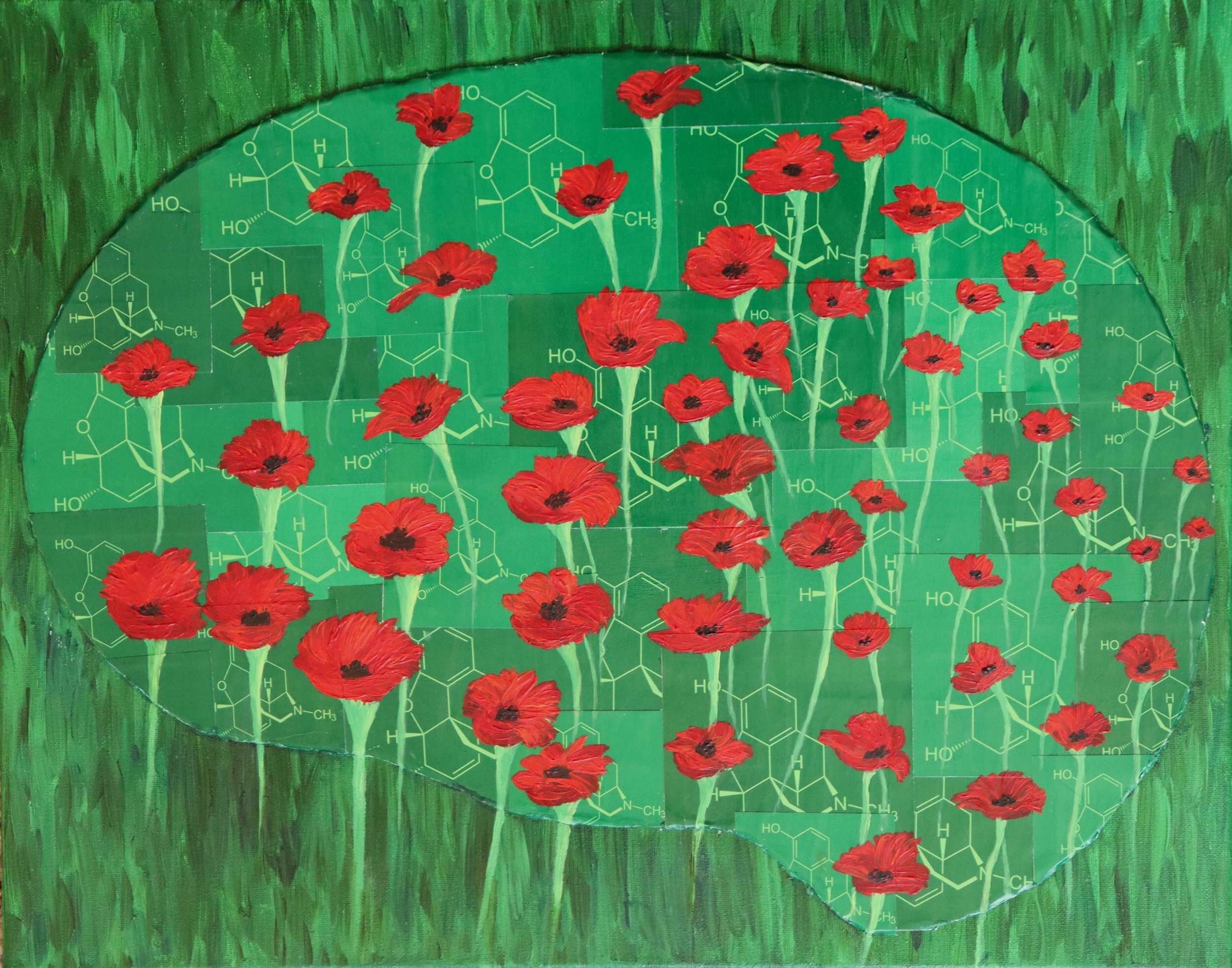
Morphine is derived from the poppy plant and used in pain control. Opioids work through receptors located across the brain and give rise to the pain-relieving effects as well as the unrelated side effects. The brain image is collaged with the chemical structure of morphine as the background and a poppy field painted across the brain.
16x20” multimedia
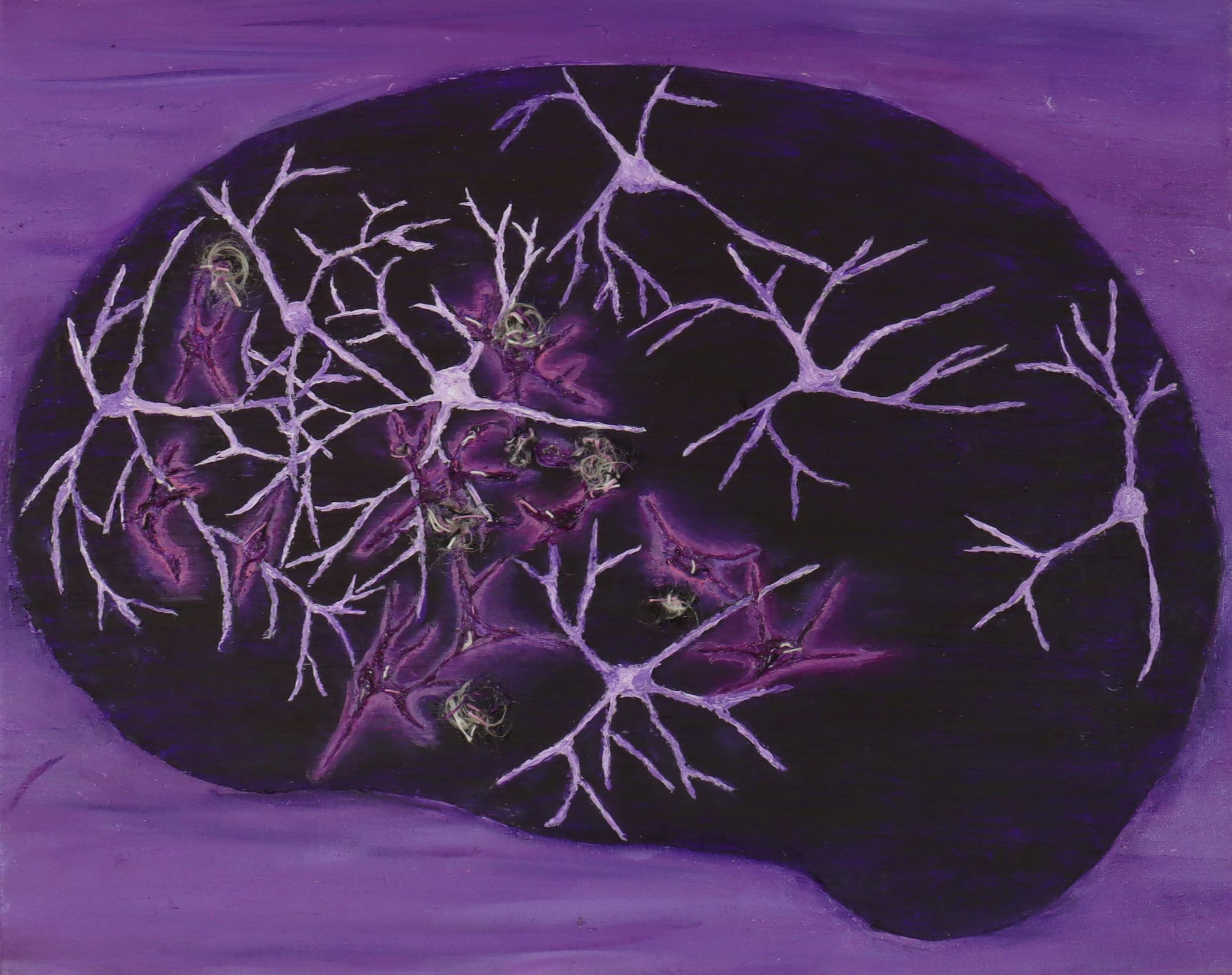
Alzheimer’s disease is a form of dementia that affects older adults leaving them with memory loss. Dying neurons (bright purple) intermingle with healthy neurons (light purple). The interfibrillary tangles and amyloid plaques are hallmarks of the disease and thought to contribute to neuron death.

Multimedia, 16x20”
SOLD
The brain is made up of complicated network of neurons. The background is collaged from National Geographic maps of the USA. Map pins represent neurons and string is used to connect neurons to show the intricate mapping of the neural circuitry.
16x20” Multimedia
SOLD
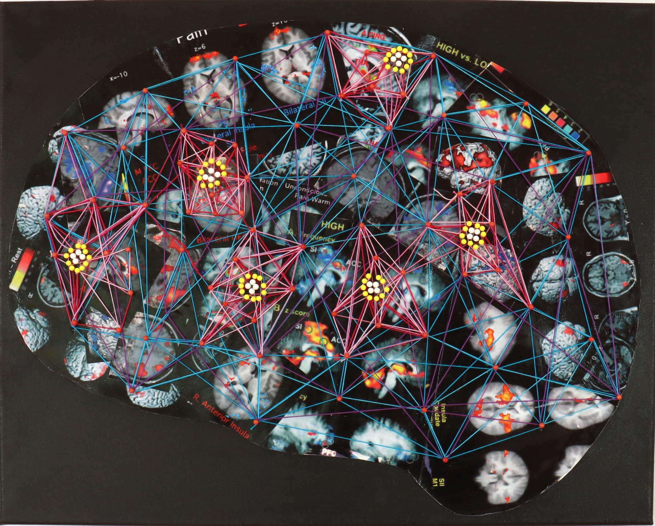
Pain is processed in a variety of brain regions. Neuroimaging techniques in human subjects has uncovered a number of brain regions involved in pain processing forming a functional map. This brain uses collaged images from scientific publications on brain imaging and pain. Map pins with brightly colored strings highlight the active regions involved in pain processing.
16x20” multimedia
SOLD
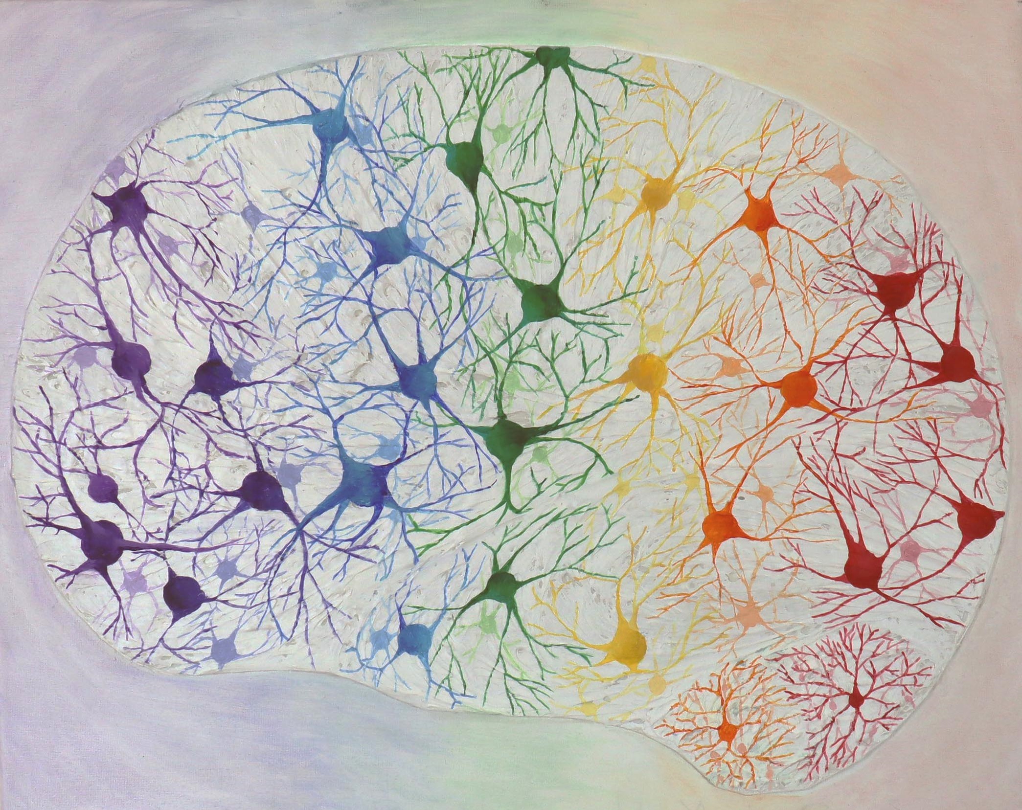
There are a variety of neurons located throughout the brain that are involved in processing signals from sensation, motor, emotions, learning, and cognitions.

16x20” Multimedia
SOLD
The brain is a series of complicated neurocircuits. Across the brain I have collaged map pins to represent neurons with colored string to represent the connections between neurons and nuclei.
16x20” Multimedia
SOLD
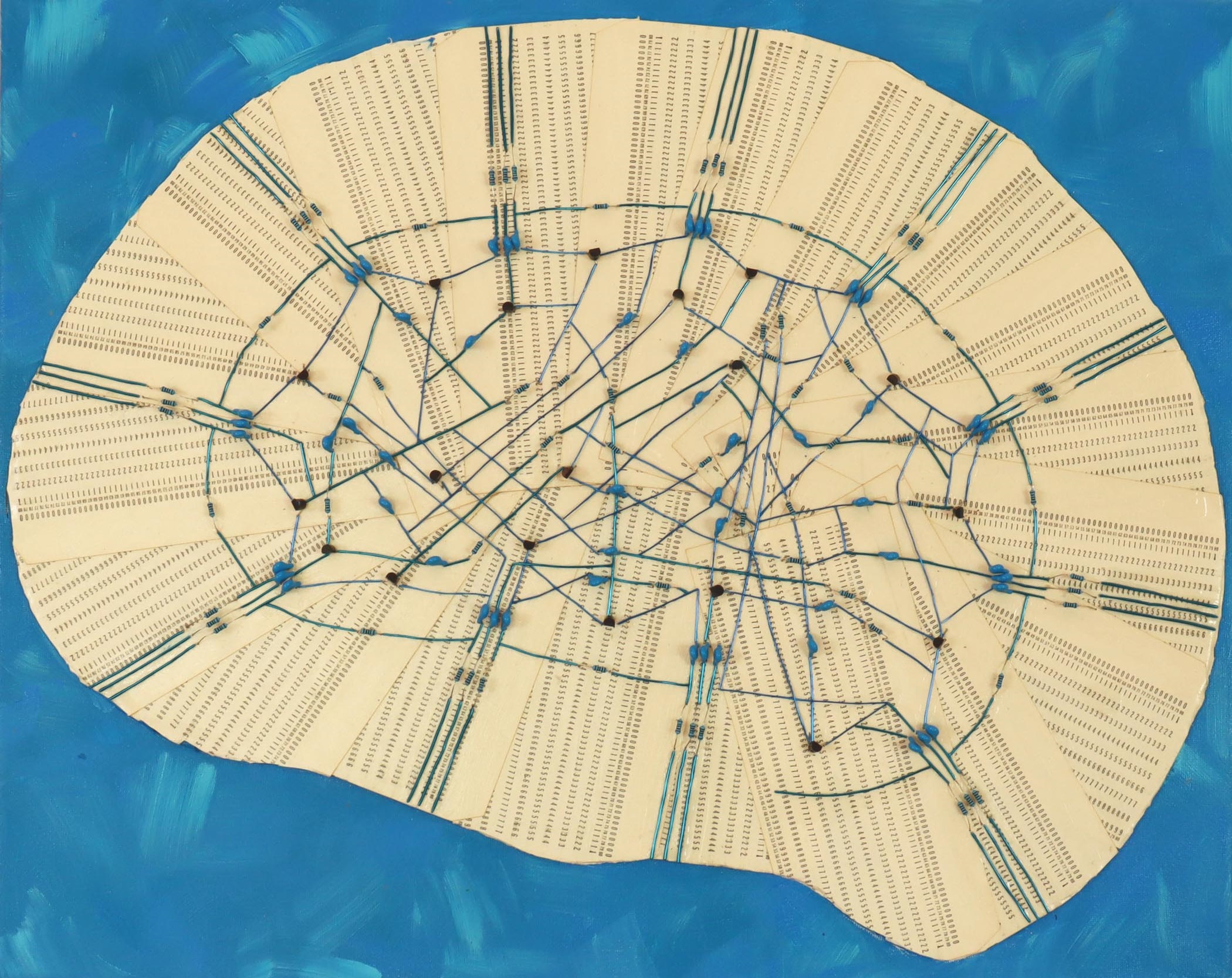
The brain has a complicated neural circuitry with more than 2 billion connections between neurons. To represent this circuitry I collaged IBM-punch cards as the background and connected brain regions using transistors, capacitors, and resistors to wires.